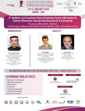
DR FODA INVITED SPEAKER IN DUBAI, UAE

Dr Foda was an invited faculty at the Emirates 10th Otorhinolaryngology Congress. Dr. Foda, joined an international Faculty of Rhinoplasty speakers including Dr. Apaydin (Turkey), Dr. Ferriera (Portugal), Dr. Durant (France), and Dr. Simmen (Swiss) in addition to some of the best regional Rhinoplasty surgeons. Dr. Foda delivered 5 lectures on different rhinoplasty topics.

Case Eight

Revision Rhinoplasty This is a 26 year old who had 3 rhinoplasties done elsewhere. On examination, the patient had a cartilaginous pollybeak, wide illdefined tip, alar asymmetry (right ala higher than left), and a short nose with an overrotated tip. Dr. Foda a revision rhinoplasty on the patient that included osteotomies (bone cuts) to narrow the nasal bones, lowering of the cartilaginous dorsum, lengthening of the nose by a caudal septum replacement graft and a double-layered tip graft, and a revision of previous columellar scar.

Case Seven

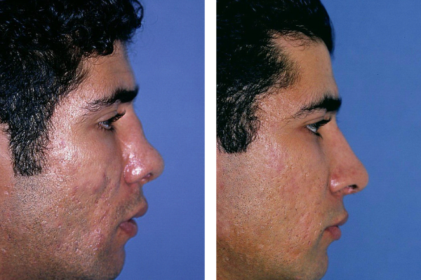
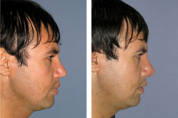
Revision Rhinoplasty This is a 34 year old male patient who had one previous nasal surgery in which most of the cartilaginous septum was excised. On examination, the patient had an external nasal deviation with depressed left nasal wall and bulging right side, cartilaginous saddle (low dorsum), depressed tip, deficient premaxilla with an acute Nasolabial angle. Dr. Foda performed a revision rhinoplasty on the patient which included osteotomies (bone cuts) to correct the bony deviation, dorsal augmentation, premaxillary augmentation with Mersilene mesh and a caudal septum replacement graft to increase the tip projection and support.

Case Six

Revision Rhinoplasty This is a 30 year old male patient who’s nose was crushed in a major car accident. The patient had two previous rhinoplasties that failed to mobilize his impacted nasal bones. On examination, the patient has a severely fractured deviated nasal bones with a traumatic saddle (concavity of nasal dorsum), superior retraction of the nasal tip resulting in an overly short nose. Dr. Foda performed a revision nasal reconstructive procedure on the patient using the patient’s own rib cartilage to build up the nasal dorsum and lengthen his short nose. Multiple osteotomies (cuts in nasal bones) were done to realign the nasal bones.

Case Five

Revision Rhinoplasty This is a 28 year old male patient who presented for revision rhinoplasty. On examination, the patient had an irregular nasal dorsum, nasal deviation to the left, and major asymmetry of the tip cartilages. Dr. Foda performed a revision rhinoplasty in which the nasal dorsum was realigned, the tip cartilages modified and sutured together in a more symmetric position.

Case Four

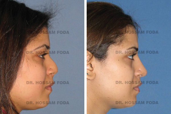
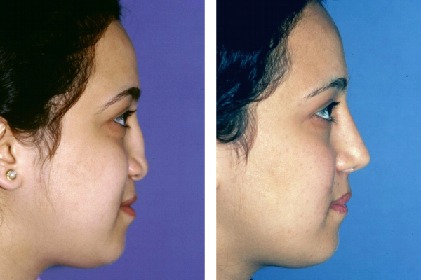
Revision Rhinoplasty This is an 18 year old female patient who presented for revision rhinoplasty. Following her original surgery, by another doctor, the patient was concerned with the appearance of her nose. On examination, the patient has an over-resected cartilaginous dorsum that resulted in a dorsal saddle on the profile view and dorsal widening on the front view. Additional finding is a droopy inferiorly rotated nasal tip due to loss of tip support. Dr. Foda performed a revision rhinoplasty on her using the patient’s ear cartilage to reconstruct the dorsal and tip support.

Case Three

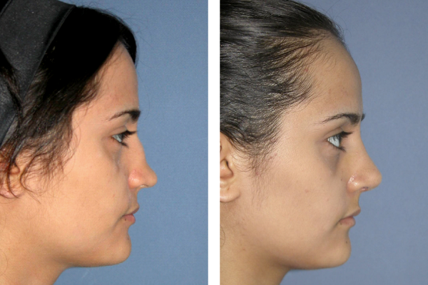
Revision Rhinoplasty This is a 20 year old female patient who had one previous unsuccessful rhinoplasty to correct her congenitally deformed nose. On examination, the patient has underdeveloped nasal cartilages with a low dorsum, depressed tip, and a very short columella (the part between the nostrils). Dr. Foda performed a revision rhinoplasty on her using the patient’s rib cartilage to build up the nasal dorsum and project the nasal tip.

Case Two

Revision Rhinoplasty This is a 25 year old male patient who had multiple failed rhinoplasties done elsewhere. The patient is complaining of bilateral nasal obstruction and of multiple nasal asymmetries. On examination, the nose shows signs of overly resected nasal cartilages in the form of lateral nasal wall depressions (more on right side). The profile view shows a retracted columella (the part between the nostrils). The basal view shows bilateral alar collapse (inwards concavity of the nostrils’ side walls). Additionally, there is marked asymmetry of alar rims (lower on the right side). Dr. Foda performed a revision rhinoplasty using the patient’s ear cartilage to restructure the nose.

Case One

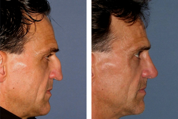
Revision Rhinoplasty This is a 47 year old male patient who had one previous nasal surgery, by another surgeon, which resulted in loss of his caudal septal cartilage with subsequently lead to displacement of the tip downwards and backwards due to the loss of tip support. Dr. Foda performed a revision rhinoplasty using a caudal septum replacement graft to resupport and reposition the nasal tip both upwards and forwards.